Medial temporal lobe (MTL) atrophy is an important morphological marker of many dementias and is closely related to cognitive decline. In this study we aimed to characterize longitudinal progression of MTL atrophy in 93 individuals with subjective cognitive decline and mild cognitive impairment followed up over six years, and to assess if clinical rating scales are able to detect these changes. All MRI images were visually rated according to Scheltens' scale of medial temporal atrophy (MTA) by two neuroradiologists and AVRA, a software for automated MTA ratings. The images were also segmented using FreeSurfer's longitudinal pipeline in order to compare the MTA ratings to volumes of the hippocampi and inferior lateral ventricles. We found that MTL atrophy rates increased with CSF biomarker abnormality, used to define preclinical stages of Alzheimer's Disease. Both AVRA's and the radiologists' MTA ratings showed a similar longitudinal trajectory as the subcortical volumes, suggesting that visual rating scales provide a valid alternative to automatic segmentations. While the MTA scores from each radiologist showed strong correlations to subcortical volumes, the inter-rater agreement was low. We conclude that the main limitation of quantifying MTL atrophy with visual ratings in clinics is the subjectiveness of the assessment.
Showing posts with label Volumetry. Show all posts
Showing posts with label Volumetry. Show all posts

Linear vs volume measures of ventricle size in hydrocephalus: relation to present and future gait and cognition
Objective To compare the clinical utility of volume-based ratios with the standard linear ratio of Evans index (EI) by examining their associations with gait, cognition, and other patient and imaging variables.
Methods From MRI scans of 1,774 participants in the Mayo Clinic Study of Aging, we calculated 3 ventricle size measures: Evan index (frontal horn width divided by widest width of skull inner table), total ventricular volume, and frontal horn volume as ratios of total intracranial volume. Gait was measured by a timed 25-foot walk and cognition by a composite of psychometric tests. We also evaluated variables associated with the measures of ventricular size. Further, we evaluated gait and cognition associations with MRI of extraventricular findings seen in normal-pressure hydrocephalus: disproportionate enlargement of subarachnoid space (DESH) and focal sulcal dilations (FSD).
Results Ventricular volume measures had stronger association with gait and cognition measures than EI. In decreasing order of strength of association with ventricle size were DESH, FSD, white matter hyperintensity volume ratio, age, male sex, cortical thickness, and education. Modest evidence was observed that FSD was associated with future decline in gait and cognition.
Conclusion Ventricular volume measures are clinically more useful than EI in indicating current and future gait and cognition. Multiple factors are associated with ventricle volume size, including FSD and DESH, suggesting that changes in CSF dynamics may go beyond simple ventriculomegaly.
DOI: https://doi.org/10.1212/WNL.0000000000008673
Thalamic nuclei in frontotemporal dementia: Mediodorsal nucleus involvement is universal but pulvinar atrophy is unique to C9orf72
Thalamic atrophy is a common feature across all forms of FTD but little is known about specific nuclei involvement. We aimed to investigate in vivo atrophy of the thalamic nuclei across the FTD spectrum. A cohort of 402 FTD patients (age: mean(SD) 64.3(8.2) years; disease duration: 4.8(2.8) years) was compared with 104 age‐matched controls (age: 62.5(10.4) years), using an automated segmentation of T1‐weighted MRIs to extract volumes of 14 thalamic nuclei. Stratification was performed by clinical diagnosis (180 behavioural variant FTD (bvFTD), 85 semantic variant primary progressive aphasia (svPPA), 114 nonfluent variant PPA (nfvPPA), 15 PPA not otherwise specified (PPA‐NOS), and 8 with associated motor neurone disease (FTD‐MND), genetic diagnosis (27 MAPT, 28 C9orf72, 18 GRN), and pathological confirmation (37 tauopathy, 38 TDP‐43opathy, 4 FUSopathy). The mediodorsal nucleus (MD) was the only nucleus affected in all FTD subgroups (16–33% smaller than controls). The laterodorsal nucleus was also particularly affected in genetic cases (28–38%), TDP‐43 type A (47%), tau‐CBD (44%), and FTD‐MND (53%). The pulvinar was affected only in the C9orf72 group (16%). Both the lateral and medial geniculate nuclei were also affected in the genetic cases (10–20%), particularly the LGN in C9orf72 expansion carriers. Use of individual thalamic nuclei volumes provided higher accuracy in discriminating between FTD groups than the whole thalamic volume. The MD is the only structure affected across all FTD groups. Differential involvement of the thalamic nuclei among FTD forms is seen, with a unique pattern of atrophy in the pulvinar in C9orf72 expansion carriers.

Related publications: Brain volume/cerebrospinal fluid index (BV/CSF index) in the diagnosis of Alzheimer's disease
BV/CSF index= (Total WM + Total GM) / Total spaces containing CSF
Background: Global brain atrophy is present in normal aging and different neurodegenerative disorders such as Alzheimer's disease (AD) and is becoming widely used to monitor disease progression. Summary: The brain volume/cerebrospinal fluid index (BV/CSF index) is validated in this study as a measurement of global brain atrophy. We tested the ability of the BV/CSF index to detect global brain atrophy, investigated the influence of confounders, provided normative values and cut-offs for mild, moderate and severe brain atrophy, and studied associations with different outcome variables. A total of 1,009 individuals were included [324 healthy controls, 408 patients with mild cognitive impairment (MCI) and 277 patients with AD]. Magnetic resonance images were segmented using FreeSurfer, and the BV/CSF index was calculated and studied both cross-sectionally and longitudinally (1-year follow-up). Both AD patients and MCI patients who progressed to AD showed greater global brain atrophy compared to stable MCI patients and controls. Atrophy was associated with older age, larger intracranial volume, less education and presence of the ApoE ε4 allele. Significant correlations were found with clinical variables, CSF biomarkers and several cognitive tests. Key Messages: The BV/CSF index may be useful for staging individuals according to the degree of global brain atrophy, and for monitoring disease progression. It also shows potential for predicting clinical changes and for being used in the clinical routine.
Reference: Camila Orellana, Daniel Ferreira, J.-Sebastian Muehlboeck, Patrizia Mecocci, Bruno Vellas, Magda Tsolaki, Iwona Kłoszewska, Hilkka Soininen, Simon Lovestone, Andrew Simmons, Lars-Olof Wahlund, Eric WestmanMeasuring Global Brain Atrophy with the Brain Volume/Cerebrospinal Fluid Index: Normative Values, Cut-Offs and Clinical Associations. Neurodegener Dis (DOI: 10.1159/000442443)
Free Supplementary Material
Free Supplementary Material
Comparison of structural MRI methodologies for measuring atrophy of structures in the medial temporal lobe
There are several approaches for quantifying the size of MTL structures using structural neuroimaging, including visual rating scales (VRS), volumetry (3D), planimetry (2D) and linear measures (1D), as depicted in this figure.

A comparison among the different methods for quantifying atrophy of the MTL based on structural neuroimaging is presented in this table.

Comparison compare the results of a manual planimetric measure (the yearly rate of absolute atrophy of the medial temporal lobe, 2D-yrA-MTL) with the results of an automated volumetric measure (the yearly rate of atrophy of the hippocampus, 3D-yrA-H).

A comparison among the different methods for quantifying atrophy of the MTL based on structural neuroimaging is presented in this table.

Comparison compare the results of a manual planimetric measure (the yearly rate of absolute atrophy of the medial temporal lobe, 2D-yrA-MTL) with the results of an automated volumetric measure (the yearly rate of atrophy of the hippocampus, 3D-yrA-H).
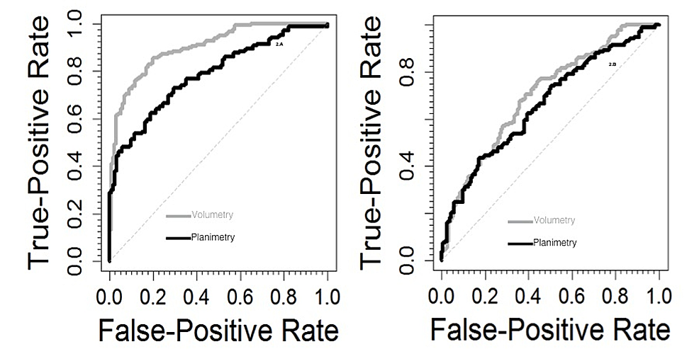 |
| ROC curves in the differential diagnosis of AD vs controls (left) and AD vs MCI (right). |
Subscribe to:
Posts (Atom)