
A comparison among the different methods for quantifying atrophy of the MTL based on structural neuroimaging is presented in this table.

Comparison compare the results of a manual planimetric measure (the yearly rate of absolute atrophy of the medial temporal lobe, 2D-yrA-MTL) with the results of an automated volumetric measure (the yearly rate of atrophy of the hippocampus, 3D-yrA-H).
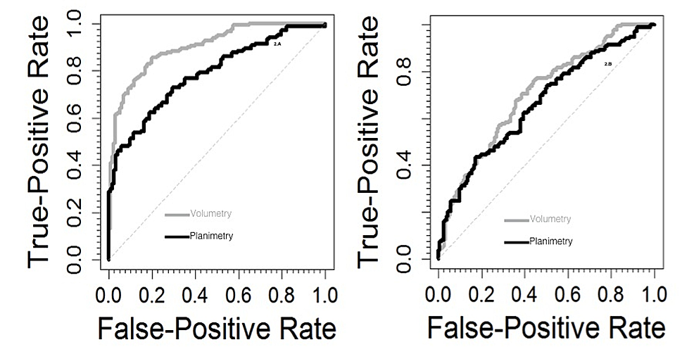 |
| ROC curves in the differential diagnosis of AD vs controls (left) and AD vs MCI (right). |
Hello. I have seen good posting. I think the table that compares the automated ratings and the visual rating is well organized. I would like to quote this table slightly modified in my article, but I would like to ask you how to apply a reference to it. Just for this site or do you have any own article to be cited? Thank you.
ReplyDeleteYes, thank you.
ReplyDeleteThat table is included in the following paper, please cite it
Manuel Menéndez-González, Aníbal Fernández Oliveira, Francisco Conejo Bayón, Jesús
Maese, Tamara Mesas Uzal, Estibaliz Herrera de la Llave, Tania Álvarez Avellón. Planimetry of the medial temporal lobe: a feasible method for supporting the diagnosis of Alzheimer’s disease in clinical practice. Neurology and Neuroscience 2015;1(8) doi: 10.3823/355
Thank you for your fast& kind reply!
DeleteThis comment has been removed by the author.
ReplyDelete